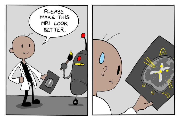
You’ve just gotten an MRI.
Most of the resulting scan is clear; however, the image quality is…suboptimal. Even with proper protocols to minimize issues, the machine, the software, and the patient’s demographics and implants can all lead to noise, artifacts and other irregularities.
Imaging in science fields is rife with such situations. From medicine to research, scientists are going to want a clear picture of their subject of interest. So, how do you solve this problem?
You use math! There are all kinds of special algorithms in use, such as image reconstruction algorithms and metal artifact reduction techniques for dealing with metal implant-induced artifacts. And with new breakthroughs in artificial intelligence, there are now new ways to boost image quality.
To learn, artificial intelligence can either use a model-free method (which relies on positive and negative reinforcement to incentivize the program towards a specific outcome) or a model-based method (which relies on using mathematical models for decision-making). Model-free methods are faster, but are less suited when more discrete traits such as age of onset of a disease are involved.
Model-based methods train by going through multiple iterations of mathematical models and using them to predict which outcome is best.
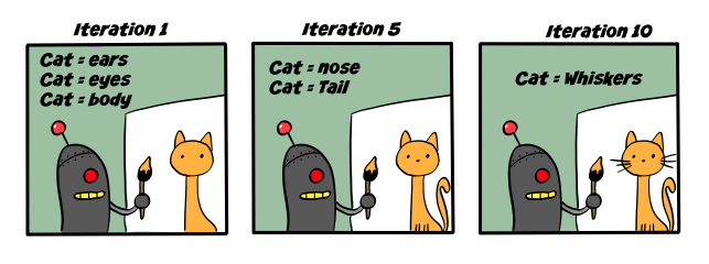
There are three strategies used for their training:
The plug-and-play method integrates pre-existing AI models and currently sees lots of use in medical imaging.
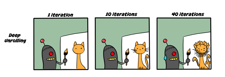
Deep unrolling methods “unroll” each iteration into different layers, and each subsequent pass through a layer improves the next layer. These methods have very high accuracy when running up to the number of iterations they have been trained on. Past that point, though, the output can get a bit…unstable.
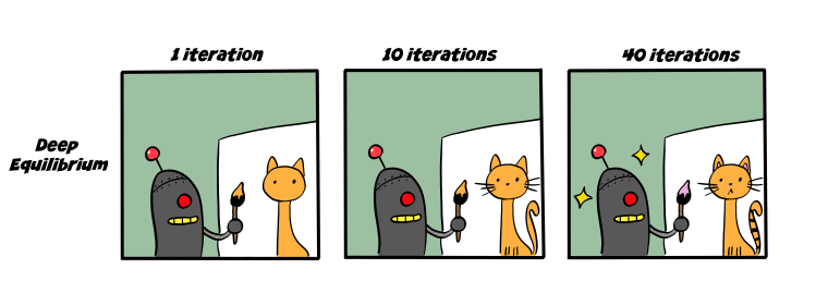
The newest and most exciting approach is the deep equilibrium method. As iterations of a deep unrolling method continue, they tend to converge towards a fixed point. By calculating that fixed point and choosing that as the endpoint, you can optimize the process by saving on memory consumption. This method is less memory-intensive and scales well to larger tasks. Gilton et al. were the first to use standard fixed-point accelerators to speed up calculation of the fixed point, further optimizing the process.
These new advances in A.I. ensure that even an imperfect MRI can yield results both useful and accurate enough for a diagnosis…as long as the model is trained properly, of course.